
Fig. 3.
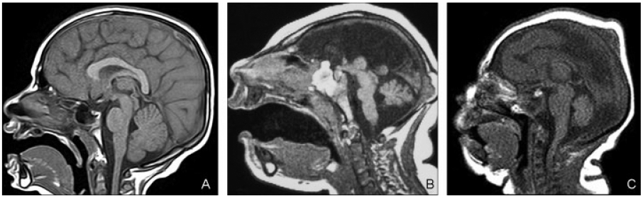
(A) Proportional cerebella. Sagittal T1-weighted image shows moderate microcephaly with proportional posterior fossa contents. (B) Proportional cerebella. A 5-day-old infant with profound microcephaly. Midsagittal T1-weighted image shows extremely small brain. Pons is short, thin, and disproportionally small compared with the cerebrum and cerebellum. Midbrain and medulla were disproportionally large compared with both cerebrum and cerebellum. (C) Proportional cerebella. Midsagittal T1-weighted image (a 1-day-old infant) shows cerebellum, midbrain, and medulla proportional to cerebrum. Pons is disproportionally large.