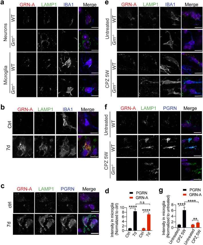
Fig. 3.
Granulin A is localized in the lysosome in neurons and microglia. a Immunostaining of GRN-A, LAMP1 and IBA1 in brain sections from 4 months old WT and Grn−/− mice. Representative images from the cortex were shown. Scale bar = 10um. b, c, d Immunostaining of GRN-A, LAMP1 and IBA1 or GRN-A, LAMP1and PGRN in brain sections from 4 months old WT mice untreated or treated with Rose Bengal and laser illumination to induce stroke (7 days after treatment). Representative images in the lesion site were shown. Scale bar = 10um. PGRN and GRN-A signals in IBA1 positive microglia were quantified by Image J. 40–50 individual microglia were quantified from multiple brain sections from 2 independent mice for each condition. Data presented as mean ± SEM. ****, p < 0.0001, n.s, not significant, unpaired two-tailed Student’s t-test. e, f, g Immunostaining of GRN-A, LAMP1 and IBA1 or GRN-A, LAMP1and PGRN in brain sections from 15-week old WT and Grn−/− mice untreated or treated with cuprizone for 5 weeks (CPZ 5 W). Representative images from the corpus callosum region were shown. Scale bar = 10um. PGRN and GRN-A signals in IBA1 positive microglia were quantified by Image J. 40–50 individual microglia were quantified from multiple brain sections from 2 independent mice for each condition. Data presented as mean ± SEM. **,p < 0.01, ****, p < 0.0001, unpaired two-tailed Student’s t-test

