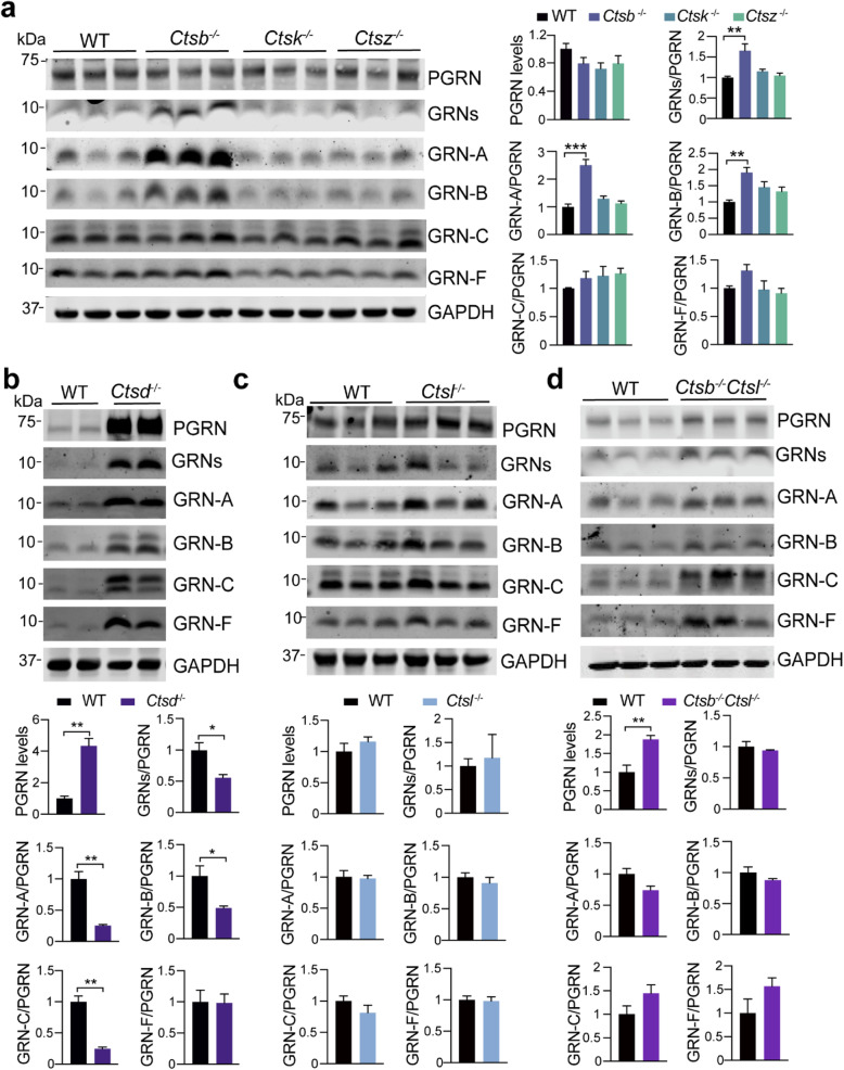
Fig. 4.
Analysis of PGRN and granulin levels in brain lysates from cathepsin deficient mice. a Western blot analysis of cortical lysates from 5 months old WT, Ctsb−/−, Ctsk−/− and Ctsz−/− mice with antibodies against each granulin peptide as indicated. The ratio between each granulin peptide to full-length PGRN was quantified and normalized to WT. Data presented as mean ± SEM. n = 3. Significant increased levels of GRNs (p = 0.0032, one-way ANOVA with Bonferroni’s post hoc comparisons), GRN-A (p = 0.0002), GRN-B (p = 0.0089) in Ctsb−/− cortical lysates compared to WT. *, p < 0.05, **, p < 0.01, ***, p < 0.001. Mixed male and female mice were used for this analysis. b-d Western blot analysis of cortical lysates from 2 weeks old Ctsd−/− mice (b), 5 months old Ctsl−/− mice (c), 3 weeks old Ctsb−/−Ctsl−/− mice (d), and age-matched WT mice using antibodies against each granulin peptide as indicated. The ratio between PGRN and GAPDH as well as between each granulin peptide and full-length PGRN was quantified and normalized to WT. Data presented as mean ± SEM. n = 3–5. The levels of PGRN are increased in Ctsd−/− (p = 0.0025) and Ctsb−/−l−/− (p = 0.0071) cortical lysates compared to WT. The ratio of total GRNs (p = 0.0286), GRN-A (p = 0.0033), GRN-B (p = 0.0373), GRN-C (p = 0.0015), but not GRN-F (p = 0.9453), versus full length of PGRN is decreased in the Ctsd−/− lysates. Data were analyzed by unpaired two-tailed Student’s t-test. *, p < 0.05, **, p < 0.01. Mixed male and female mice were used for this analysis

