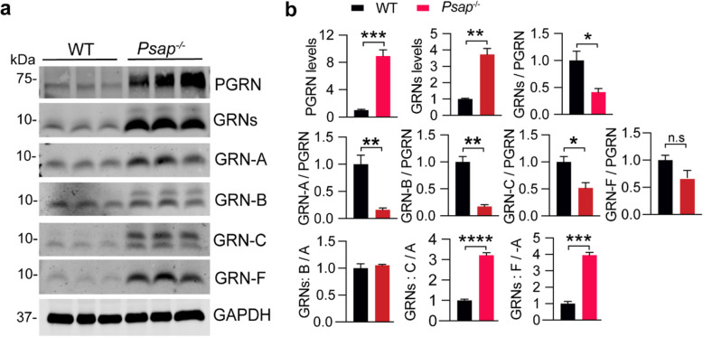
Fig. 5.
Analysis of PGRN and granulin levels in brain lysates from PSAP deficient mice. a, b Western blot analysis of cortical lysates from 3-week old WT and Psap−/− mice with antibodies against each granulin peptide and full-length PGRN as indicated. The ratio between PGRN or total granulins or each granulin peptide and GAPDH as well as between granulins and full-length PGRN was quantified and normalized to WT. The ratios between granulins B/C/E/F and granulin A were quantified and shown in (b). Data presented as mean ± SEM. n = 3. The levels of PGRN (p = 0.0009) and granulins (p = 0.0017) are increased in Psap−/− mouse cortical lysates compared to WT. The ratio of total GRNs (p = 0.0339), GRN-A (p = 0.0078), GRN-B (p = 0.0016), GRN-C (p = 0.025), but not GRN-F (p = 0.093), versus full length of PGRN is decreased in the Psap/− lysates. Data were analyzed by unpaired two-tailed Student’s t-test. *, p < 0.05, **, p < 0.01, ***, p < 0.001. Mixed male and female mice were used for this analysis

