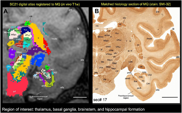
Fig. 14. Registration of subcortical atlas to a different test subject with histological confirmation of architectonic areas.
In this example, the segmented subcortical 3D volume (SC21) is registered to the T1w MRI volume of a different individual brain (Case MQ). (A) Shows the registered subcortical areas from SC21 overlaid on the MQ coronal MRI slice. None of the registered regions were altered or adjusted in this MRI slice or 3D volume. (B) Matched histology section from MQ stained immunohistochemically for the neurofilament protein, recognized by SMI-32 antibody (sec# 17). For consistency we flipped both MRI and histology in this illustration (i.e., left hemisphere is on the right). We digitally rotated the T1w MRI volume to match with the histology sections before the registration process. Note the correspondence of sulci and gyri in both the registered volume and histology section. We also confirmed the spatial location and the architectonic features of the selected subcortical targets (subregions of the thalamus, basal ganglia, brainstem, and hippocampal formation) in the registered coronal slice with the corresponding histology section as illustrated in A and B. For more details on the subregions of these deep brain structures see the result section. Abbreviations-Thalamus: cl-central lateral nucleus; cnMD-centromedian nucleus; LD-lateral dorsal nucleus; LGN-lateral geniculate nucleus; MDmc-medial dorsal nucleus, magnocellular division; MDmf-medial dorsal nucleus, multiform division; MDpc-medial dorsal nucleus, parvicellular division; Pf-parafascicular nucleus; r-reticular nucleus; VLc-ventral lateral caudal nucleus; VPI-ventral posterior inferior nucleus; VPLc-ventral posterior lateral caudal nucleus; VPLo-ventral posterior lateral oral nucleus; VPM-ventral posterior medial nucleus; VPMpc-ventral posterior medial nucleus, parvicellular division; zic-zona inserta. Basal ganglia and related fiber tracts: al-ansa lenticularis; cd-caudate nucleus; cdt-caudate tail; ic-internal capsule; lf-lenticular fasciculus; mb-Muratoff bundle; pu-putamen; SNpc: substantia nigra, pars compacta; SNpl: substantia nigra, pars lateralis; SNpm: substantia nigra, pars mixta; SNpr: substantia nigra, pars reticulata. Brainstem structures: CP-cerebral peduncle; IPN-interpeduncular nucleus; pcf-pontocerebellar fibers; PN-pontine nuclei; RN-red nucleus; VTA-ventral tegmental area. Hippocampal formation: CA1-CA4-subfields of the hippocampus; FD-fascia dentata; paraS-parasubiculum; preS-presubiculum; proS-prosubiculum; Sub-subiculum. Other structures: CC-corpus callosum; f-fornix. Sulci: ips-intraparietal sulcus; ls-lateral sulcus; ots-occipitotemporal sulcus; sts-superior temporal sulcus. Scale bar: 5 mm (A and B).

