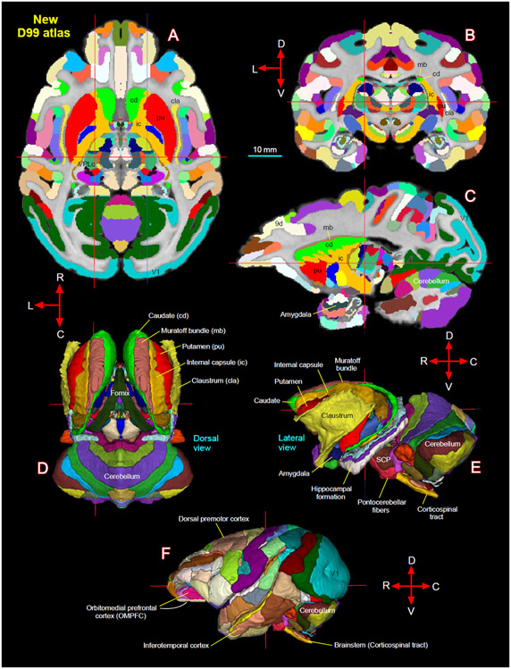
Fig. 15.
[A-C] New D99 digital atlas (version 2.0) with combined cortical and subcortical segmentation overlaid on the horizontal, coronal, and sagittal D99 ex-vivo MRI template. The cross-hairs in A-C show the same location of thalamic subregion VPLc (ventral posterior lateral caudal nucleus). (D-E) The spatial location of segmented subcortical regions shown on the dorsal and lateral views in 3D. The selected subcortical regions in D-E are also indicated with cortical areas in A-C. (F) Segmentation of cortical areas. Abbreviations: 9d-dorsal prefrontal area; SCP-superior cerebellar peduncle; V1-primary visual cortex. Orientation: D-dorsal; V-ventral; R-rostral; C-caudal; L-lateral. Scale bar: 10 mm applies to A-C only.

