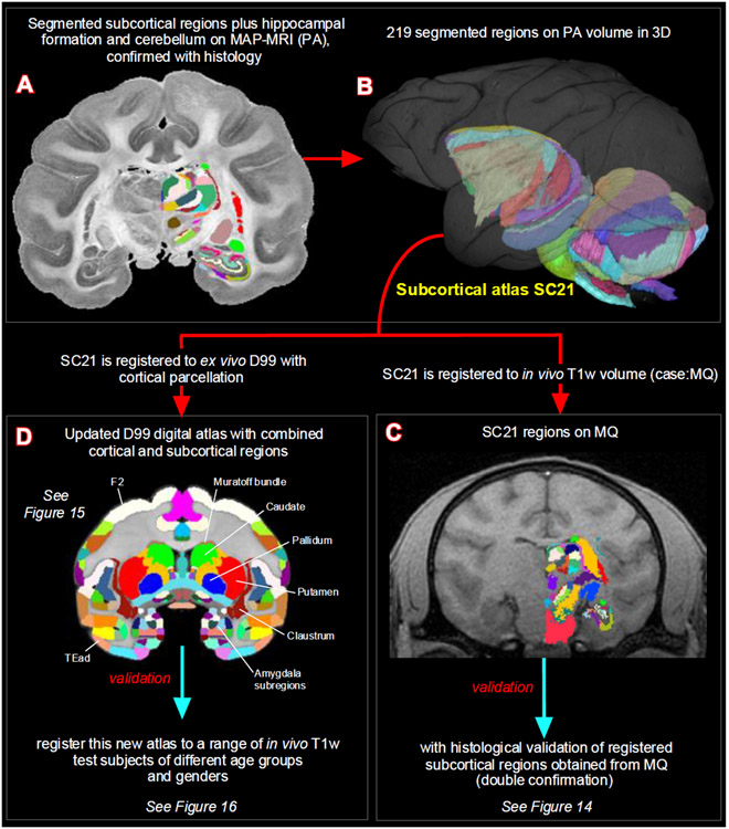
Fig. 2. Subcortical segmentation, updated 3D digital template atlas, and registration of 3D atlas to test subjects.
A total of 219 deep brain regions, including the hippocampal formation and the cerebellum were manually segmented through a series of 200 μm thick MAP-MRI sections (A) using ITK-SNAP and derived the spatial location of these regions in 3D (B). This new MRI-histology based segmented volume (called “SC21”) is registered to in vivo T1-weighted MRI volume of a test subject MQ (C). The registered subcortical regions on MQ are confirmed again using the corresponding histological sections from the same test subject. The SC21 is also registered to the standard D99 cortical template atlas (Reveley et al., 2017), resulting in an “updated D99 atlas” with combined cortical and subcortical parcellations in the same volume (D). This updated D99 is in turn registered to in vivo T1-weighted MRI volumes of 6 other subjects with different age groups. For more details, see figures 14-16.

