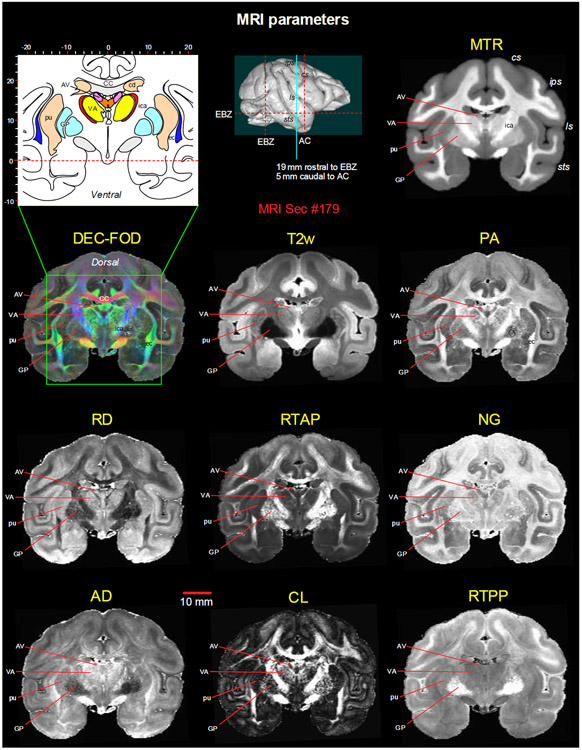
Fig. 3. Subcortical regions in different MRI parameters.
A coronal MR image from eight DTI/MAP-MRI parameters, T2-weighted (T2w), and the magnetization transfer ratio (MTR) images show four selected subcortical regions: two thalamic nuclei (AV and VA), and two basal ganglia regions (pu and GP). These areas are also illustrated in the corresponding drawing of the section on the top left (inset). This MRI slice is located 19 mm rostral to the ear bar zero (EBZ) or 5 mm caudal to the anterior commissure (AC) as illustrated by a blue vertical line on the lateral view of the 3D rendered brain image. Note that the contrast between these subcortical areas is distinct in different MRI parameters. Abbreviations: DTI/MAP-MRI parameters: AD-axial diffusivity; CL-linearity of diffusion from the tortoise; DEC-FOD-directionally encoded color-fiber orientation distribution; NG-non-gaussianity; PA-propagator anisotropy; RD-radial diffusivity; RTAP-return to axis probability; RTPP-return to plane probability. Subcortical regions: AV-anterior ventral nucleus; ec-external capsule; ica-internal capsule, anterior limb; VA-ventral anterior nucleus; GP-globus pallidus; pu-putamen. Sulci: cs: central sulcus; ips-intraparietal sulcus; ls-lateral sulcus; sts-superior temporal sulcus.

