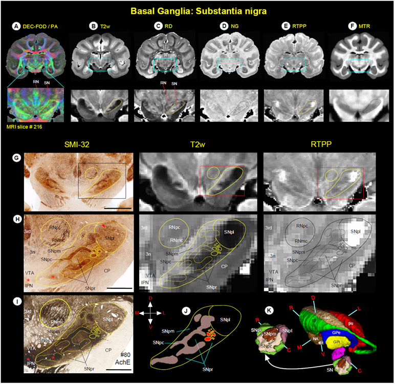
Fig. 6.
(A-F) Zoomed-in view of the green boxed region shows the spatial location of substantia nigra (SN), and the adjacent red nucleus (RN) in coronal MAP-MRI (DEC-FOD, RD, NG, RTPP), T2w, and MTR images. (G-J, left and right columns) The high-power photographs of the SMI-32 and AchE stained sections, and T2w and RTPP show four subregions of the SN: substantia nigra- pars compacta (SNpc), pars lateralis (SNpl), pars mixta (SNpm), and pars reticulata (SNpr). In addition, H and I also show the transition zone of the parvicellular and magnocellular subregions of the RN (RNpc and RNmc, respectively). The solid and dashed outlines on the MRI depicting the subregions are derived from histology section #80 on the left. (K) The spatial location and overall extent of the different subdivisions of the SN with other subregions of the basal ganglia in 3D, reconstructed using ITK-SNAP. For the abbreviations of basal ganglia regions, see figure legends 4 and 5. Orientation: D-dorsal; V-ventral; R-rostral; C-caudal; M-medial; L-lateral. Abbreviations: 3rd-oculomotor nucleus; 3n-oculomotor nerve; CP-cerebral peduncle; IPN-interpeduncular nucleus; VTA-ventral tegmental area. Scale bar: 5 mm (G) and 2 mm (H, I).

