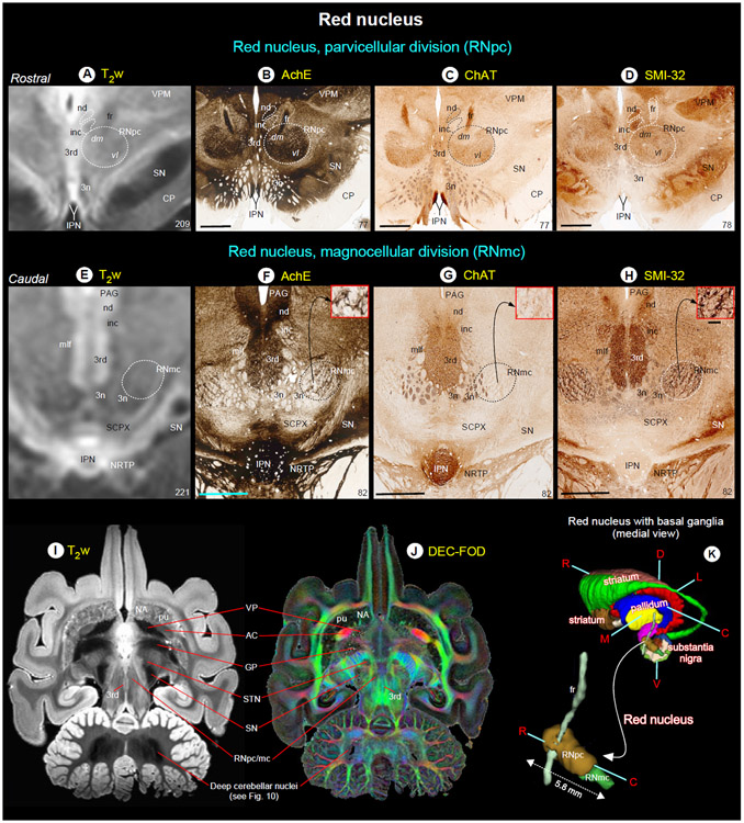
Fig. 7. Red nucleus.
(A-H) show the rostral parvicellular (RNpc) and the caudal magnocellular (RNmc) subregions of the red nucleus in coronal T2w, and matched histological sections. Note that the borders of RNpc and RNmc from the surrounding subcortical regions are less prominent in T2w than in AchE, ChAT and SMI-32 stained sections. The dashed outlines on the T2w images are reproduced from corresponding regions in the histology images. The numbers on the bottom right indicate the MRI slice number and histology section number. Other prominent gray and white matter regions surrounding the red nucleus are visible in these images (see abbreviations below). (I-J) illustrate the signal intensity differences between RN and subregions of the basal ganglia in horizontal T2w and MAP-MRI (DEC-FOD) images. In contrast to red nucleus (RNpc/mc), the globus pallidus (GP), ventral pallidum (VP), and substantia nigra (SN) exhibited significantly decreased (hypointense) signal in T2w image (see also deep cerebellar nuclei), probably due to the high level of iron content. These subregions show variable signal intensities in DEC-FOD image as shown in J. (K) 3D reconstruction shows the spatial location of the subregions of red nucleus with reference to basal ganglia and fr, a fiber tract which pierce through the RNpc. Abbreviations: 3rd-oculomotor nuclei; 3n-oculomotor nerve; AC-anterior commissure; CP-cerebral peduncle; fr-fasciculus retroflexus; inc-interstitial nucleus of Cajal; IPN-interpeduncular nucleus; mlf-medial longitudinal fasciculus; nd-nucleus of Darkschewitsch; NRTP-nucleus reticularis tegmenti pontis; PAG-periaqueductal gray; SCPX-superior cerebellar peduncle decussation; STN-subthalamic nucleus; VPM-ventral posterior medial nucleus. Orientation: D-dorsal; V-ventral; R-rostral; C-caudal; M-medial; L-lateral. Scale bars: 2 mm applies to all histology images; 0.25 mm applies to inset in H.

