Figure 7.

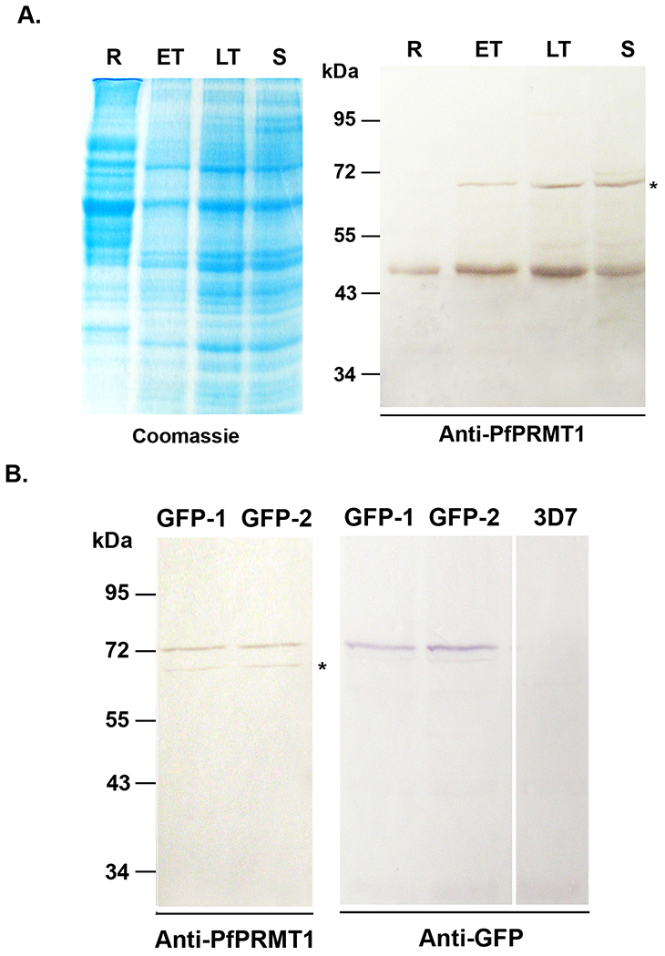
PfPRMT1 expression in the parasite. A. Time course expression of PfPRMT1 during IDC. Equal amounts of parasite lysate (~30 μg) were separated in 10% SDS-PAGE and probed with polyclonal anti-PfPRMT1 antiserum. Left panel is the Coomassie blue-stained gel to indicate approximately equal loading. Asterisk indicates the cross-reacting protein. R – ring, ET – early trophozoite, LT – late trophozoite, S – schizont. B. Confirmation of the C-terminal GFP tagging of the endogenous PfPRMT1 locus. Two clones (GFP-1 and GFP-2) were probed with the anti-PfPRMT1 antiserum (left panel) or the anti-GFP antibody (right panel). Lysate from wild-type 3D7 parasite was included as a GFP-negative control.
