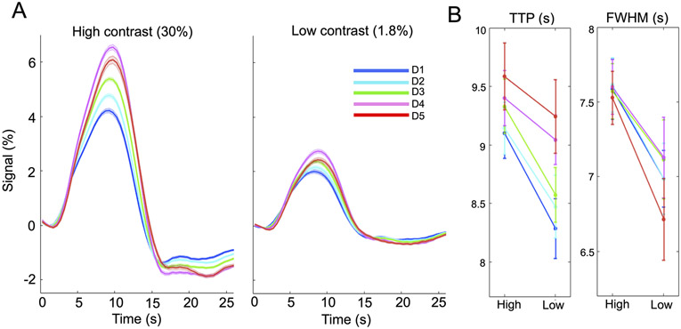
Figure 2.
Contrast-dependent fMRI patterns across cortical depths (6-s trial case, task-active voxels with F-score > 10). (A) Cortical-depth-dependent HRFs evoked by lower and higher luminance levels (mean and standard errors across subjects), with depths D1–D5 defined in 2.1.1.4; (B) Reduced TTPs and FWHMs of lower-contrast HRFs. Contrast-dependent TTP and FWHM alterations remained if the F-score threshold was varied from 5 to 15, except that in a few subjects’ results, the percent signal change of the above-pial HRF (‘red’) became smaller than those in deeper depths when we lowered the F-score threshold.

