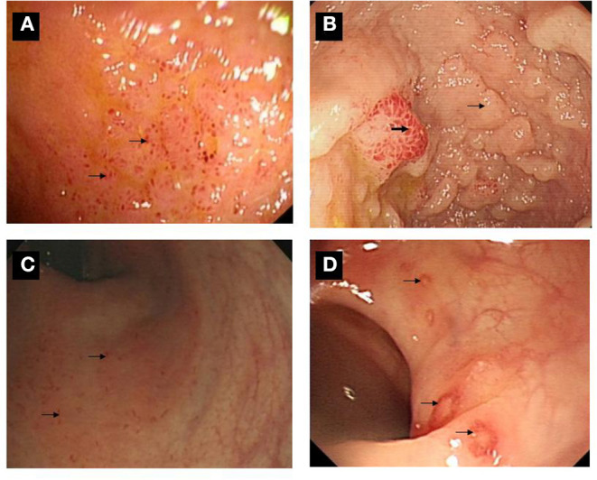
Figure 3.

Ileo-colonoscopic pictures of gut inflammation among SpA patients. (A) Multiple lesions were over the surface in the terminal ileum. (B) A sign of mucosal nodularity (small arrow) with hyperemia-like changes was in terminal ileum (bold arrow). (C) Multiple small lesions were found over the surface of rectum and sigmoid colon (arrow). (D) Multiple ulcers were found over the surface of descending, sigmoid colon and rectum (arrow).
