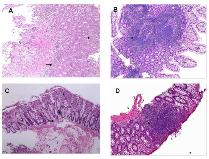
Figure 4.
Histopathological pictures of gut inflammation among SpA patients. (A) Histologically infiltration of Inflammatory cells was around gland duct (small arrow) and at underlying submucosa (bold arrow) located in terminal ileum (H&E; original magnification ×10). (B) Multiple ectopic lymphoid tissues were microscopically observed in the terminal ileum (long arrow) with massive infiltration of inflammatory cells around gland duct (H&E; original magnification ×10). (C) Tissue specimen from descending colon demonstrated moderate infiltration of inflammatory cells around gland duct (small arrow) and in the underlying submucosa (bold arrow) (H&E; original magnification ×20). (D) A ectopic lymphoid tissue was microscopically observed at rectum specimen (long arrow) with massive infiltration of inflammatory cells around gland duct (H&E; original magnification ×20).

