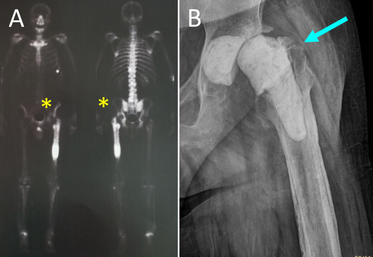
Figure 1. PJI of the left hip of a 65-year-old man with Staphylococcus aureus-infected left THR.
A. 99m Tc bone scan shows increased radioisotope uptake at the proximal metaphysis and the diaphysis of the left femur (yellow asterisks). B. Anteroposterior radiograph of the left hip after removal of the prosthesis, extensive debridement and implantation of a ball-and-socket ALCS (blue arrow).
PJI: prosthetic joint infection, THR: total hip replacement, ALCS: antibiotic-loaded cement spacer

