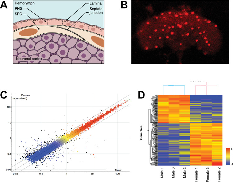
Fig 1. Microarray analysis of isolated SPG cells of the BBB.
(A) Schematic of the Drosophila Blood Brain Barrier (BBB). The BBB consists of two layers of glial cells, the outer Perineurial Glia (PG) facing the circulating hemolymph, and the inner Subperineurial Glia (SPG) with septate junctions that form the main barrier. The SPG is in contact with the underlying nuclei of the neuronal cortex. (B) Isolated fly brain with SPG cells labeled by nuclear DsRed expression driven by SPG-Gal4. Fluorescently marked cells like these from males and females were hand-isolated for RNA extraction. 20x Magnification. (C) Probes present (above background) in all male or female samples are displayed as normalized to the 75th percentile intensity of each array (19,218 probes). Each spot is the mean of 3 samples from each condition. Black spots = differentially expressed genes (>2Fold, T-test p-value < 0.05, 284 probes). Red/orange = High expression, Yellow = Medium expression, Blue = Low expression. (D) Differentially expressed genes (>2 fold, T-test p-value < 0.05) in Male vs. Female are displayed as normalized to the median value of each probe across six samples (284 probes). The heat map color scale is shown on the right.

