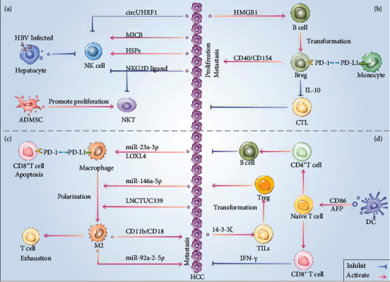
Figure 4.

The landscape of tumor Immune microenvironment regulated by exosomes in HCC. (a) Exosomes containing specific cargos activate the function of NK cell or inhibit the function of both NK cell and NKT cell, while the function of NK cell can be inhibited by exosomes secreted by HBV infected hepatocyte and the proliferation of NKT cell can be boosted by exosomes from ADMSC. (b) Breg cells either release IL-10 to inhibit the function of CTL, which participates in reversing the progression of HCC, or express CD40/CD154 to directly stimulate the proliferation and metastasis of HCC cells, whereas PD-L1 expressed on monocytes combined with PD-1 on Breg cells lead to the exhaustion of Breg cell. (c) Macrophage expresses a higher level of PD-L1 resulting from incorporating exosomes derived from HCC, later interacting with PD-1 on CD8+T cell, and luring the apoptosis of CD8+T cell. And the same exosomes but different cargos trigger the polarization of the macrophage into the M2 subtype. (d) Exosomes containing CD86/AFP derived from DC activate naïve T cells into CD4+ T cell or CD8+T cell. Meanwhile, exosomes from HCC containing 14-3-3ζ transform TILs into Tregs, thus formatting the immunosuppressed environment of HCC.
