
Figure 5.
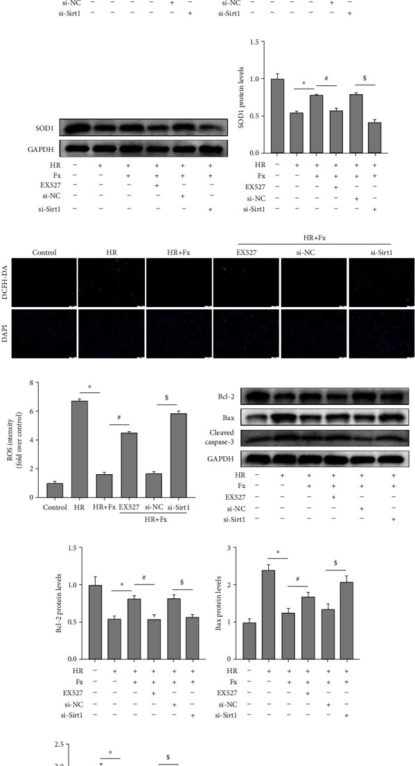
Inhibition of Sirt1 reversed the role of fucoxanthin in protecting HK-2 cells from H/R injury. The H/R model was established by hypoxia for 12 hours and reoxygenation for 6 hours. The HK-2 cells were treated with 5 μM fucoxanthin, 20 μM EX527, si-NC, or si-Sirt1 for 24 hours and then experienced H/R. (a)–(d) Sirt1, Nrf2, and HO-1 protein expression and their quantitative analysis. (e) and (f) SOD1 protein expression and its quantitative analysis. (g) and (h) Representative images of DCFH-DA staining (magnification ×100; scale bars = 100 μm) and their quantitative analysis. (i)–(l) Expression of apoptosis-related proteins Bcl-2, Bax, cleaved caspase-3, and their quantitative analysis. (m) and (n) Percentage of apoptotic cells detected by flow cytometry. Values are expressed as mean ± SEM, n = 3.∗p < 0.05 compared with HR group, #p < 0.05 compared with HR + Fx group, $p < 0.05 compared with HR + Fx + si − NC group.