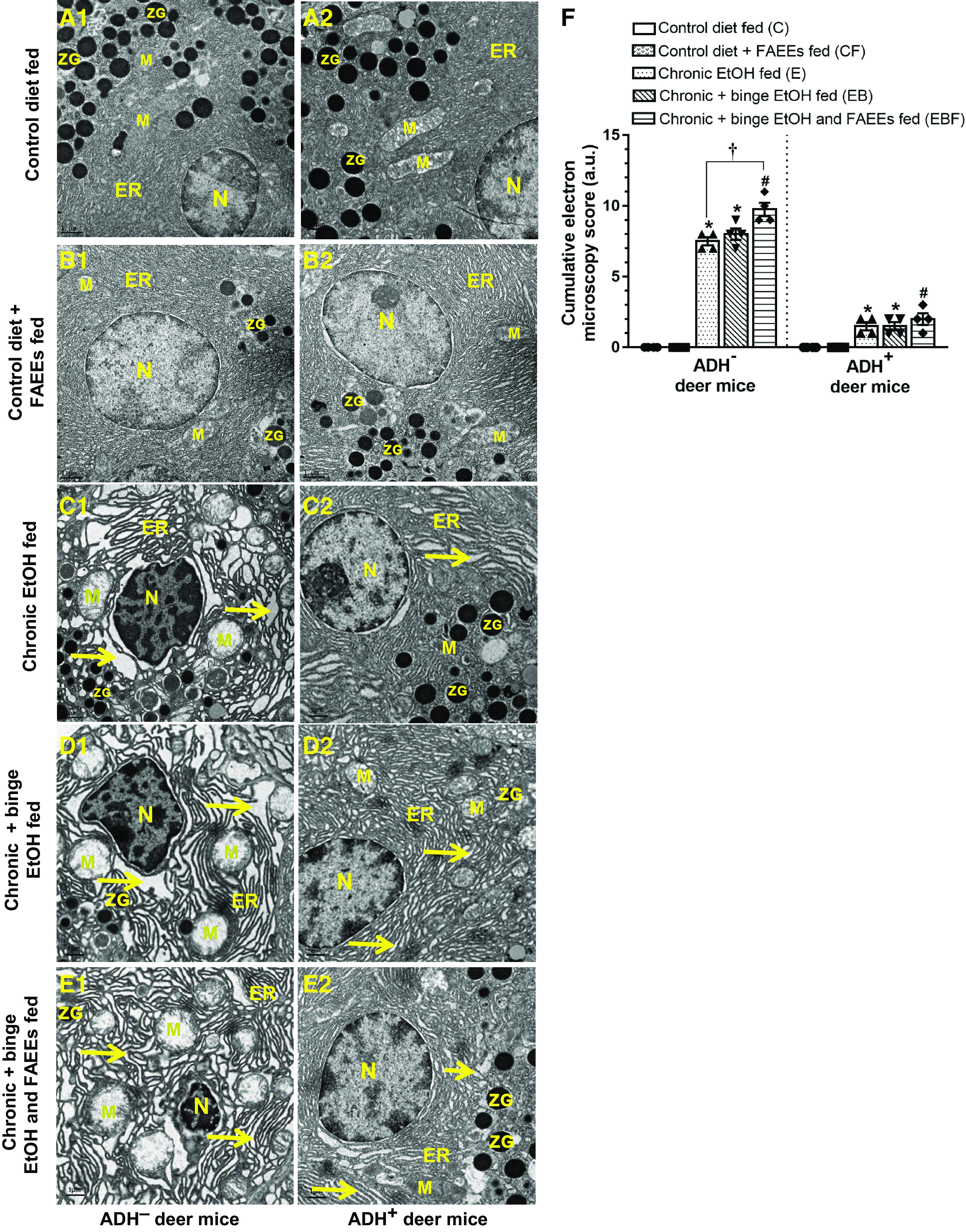
Figure 4.

Electron micrographs of pancreatic sections of ADH− (A1–E1; left) and ADH+ deer mice (A2–E2; right). (Scale bars = 1 µm). Pancreas of pair-fed control and control diet and FAEEs-fed ADH− (A1 and B1) and ADH+ (A2 and B2) deer mice show normal ultrastructure with intact nucleus mitochondria and ER cisteranes. Pancreas of chronic EtOH-fed (C1), chronic plus binge EtOH-fed (D1), and chronic plus binge EtOH and FAEEs-fed (E1) ADH− deer mice shows extensive dilatations of ER cisternae along with swollen mitochondria with short and broken cristae and shrunken nucleus. Pancreas of chronic EtOH-fed (C2), chronic plus binge EtOH-fed (D2), and chronic plus binge EtOH and FAEEs-fed (E2) ADH+ deer mice show mild to moderate dilatations in ER cisteranes with normal and intact mitochondria and nucleus. Cumulative electron microscopy score in the pancreatic ultrastructure of ADH− and ADH+ deer mice (F). Arrows indicate the dilatation of ER cisternae. Data were analyzed using ANOVA, followed by Tukey’s multiple comparisons test, and presented as means ± SE (n = 4 replicates). *P ≤ 0.05 chronic EtOH-fed group and chronic plus binge EtOH-fed group vs. pair-fed control diet group. #P ≤ 0.05 chronic plus single binge EtOH and FAEEs-fed group vs. control diet plus FAEEs-fed group and †P ≤ 0.05 chronic plus binge EtOH and FAEEs-fed group vs. chronic EtOH-fed group, respectively. ADH−, alcohol dehydrogenase-deficient; ER, granular endoplasmic reticulum; FAEEs, fatty acid ethyl esters; M, mitochondria; N, nucleus; ZG, zymogen granules.
