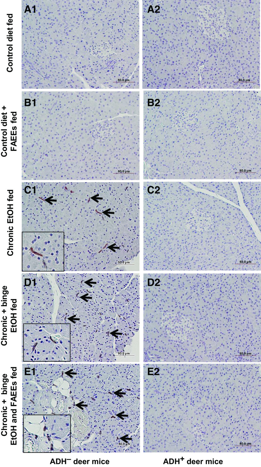
Figure 6.
Immunohistochemical staining using antibodies against F4/80 (macrophage marker) in representative pancreatic sections of ADH− (A1–E1; left) and ADH+ (A2–E2; right) deer mice. Macrophages or positive staining for F4/80 were found in the exocrine parenchyma of various chronic EtOH-fed groups of ADH− deer mice (C1–E1) as compared with ADH+ deer mice (C2–E2), respectively. No positive staining for F4/80 was observed in the pancreas of pair-fed control diet and control diet plus FAEEs-fed ADH− (A1 and B1) and ADH+ (A2 and B2) deer mice, respectively. Arrows represents positive staining for F4/80. Inset shows area of higher magnification (×40). ADH−, alcohol dehydrogenase-deficient; FAEEs, fatty acid ethyl esters.

