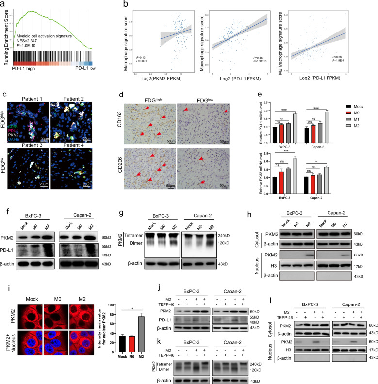
Fig. 3. M2 macrophages upregulate PD-L1 expression in tumor cells by inducing nuclear translocation of PKM2 in PDAC.
a GSEA of myeloid cell activation signature in PD-L1hi PDAC samples vs. PD-L1lo counterparts from the TCGA dataset. b TCGA analysis between the scoring of macrophage signature and PKM2 (left) or PD-L1 (middle). Also, correlation between M2 macrophage signature and PD-L1 was calculated in PDAC (right). c Representative immunofluorescence staining of CD68+ macrophages (green), PD-L1+ cells (red), and PKM2+ cells (white) in PDAC tissues with FDGhi or FDGlo. Scale bar, 20 μm. d Representative immunostaining of CD163+/CD206+ macrophages from paired tumors of patients with PDAC with FDGhi or FDGlo. Red arrowheads showed CD163+ or CD206+ macrophages. Scale bar, 50 μm. e BxPC-3/Capan-2 cells were co-cultured with M0, M1, or M2 polarized macrophages (tumor cell:macrophage = 1:2) for 48 h. Then, BxPC-3/Capan-2 cells were lysed and assayed for the expression of PD-L1 and PKM2 mRNA by qRT-PCR. f PKM2 and PD-L1 protein expression levels from whole-cell lysate of BxPC-3/Capan-2 cells co-cultured with M0/M2 macrophages were determined by western blotting. g BxPC-3/Capan-2 cells co-cultured with M0/M2 macrophages were cross-linked by glutaraldehyde first and then subjected to western blotting. h Nuclear and cytosolic lysates were prepared from BxPC-3/Capan-2 cells co-cultured with M0/M2 macrophages, followed by western blotting. i Subcellular localization of PKM2 in BxPC-3/Capan-2 cells co-cultured with M0/M2 macrophages. Cells were immunostained with anti-PKM2 (PKM2, red). The nucleus was marked with 4′,6-diamidino-2-phenylindole dihydrochloride (blue). j BxPC-3/Capan-2 cells were pretreated with TEPP-46 (10 μM) for 12 h or/and then co-cultured with M2 polarized macrophages (tumor cell:macrophage = 1:2) for 48 h. Then, PKM2 and PD-L1 protein expression levels from whole-cell lysate of BxPC-3/Capan-2 cells were determined by western blotting. k BxPC-3/Capan-2 cells pretreated with TEPP-46 or/and co-cultured with M2 macrophages were cross-linked by glutaraldehyde first and then subjected to western blotting. l Nuclear and cytosolic lysates were prepared from BxPC-3/Capan-2 cells pretreated with TEPP-46 or/and co-cultured with M2 macrophages, followed by western blotting. Data were shown as mean ± SEM from three experiments. Statistical significance was determined by a t-test; *P < 0.05,**P < 0.01,***P < 0.001. NS = no significance.

