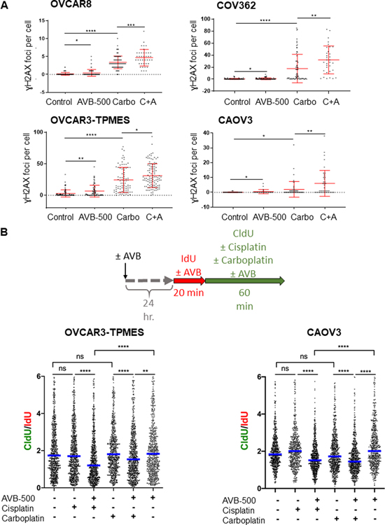
Figure 4. AVB-500 alone or in combination with chemotherapy increases DNA damage and slows replication fork progression.
A, Number of γH2AX foci per nucleus induced by treatment with vehicle, 1 μM AVB-500, 500 μM carboplatin (Carbo), or carboplatin plus AVB-500 (C+A) in OVCAR8, COV362, OVCAR3-TPMES, and CAOV3 cells. Cells were treated for 4 hours. Foci were quantified from two technical replicates with n>100 cells per experiment. Error bars indicate ± SD. *P<0.05, **P<0.01, ***P<0.001, ****P<0.0001 by student’s two-tailed t-test. B, Top, Schematic for DNA fiber assays. Bottom, CldU/IdU ratios in OVCAR3-TPMES cells ± 1 μM AVB-500 ± 150μM cisplatin ± 500 μM carboplatin (n=3) and CAOV3 cells ± AVB-500 ± cisplatin ± carboplatin (n=2) cells treated as indicated. Horizontal lines indicate medians. CldU was added concomitantly with 150 μM cisplatin or 500 μM carboplatin. In the case of AVB-500 treatment, cells were treated with 1 μM AVB-500 24 hours before the start of the fiber assay, and 1 μM AVB-500 was maintained in the cell media during the entire labeling period. **P < 0.01, **** P < 0.0001.

