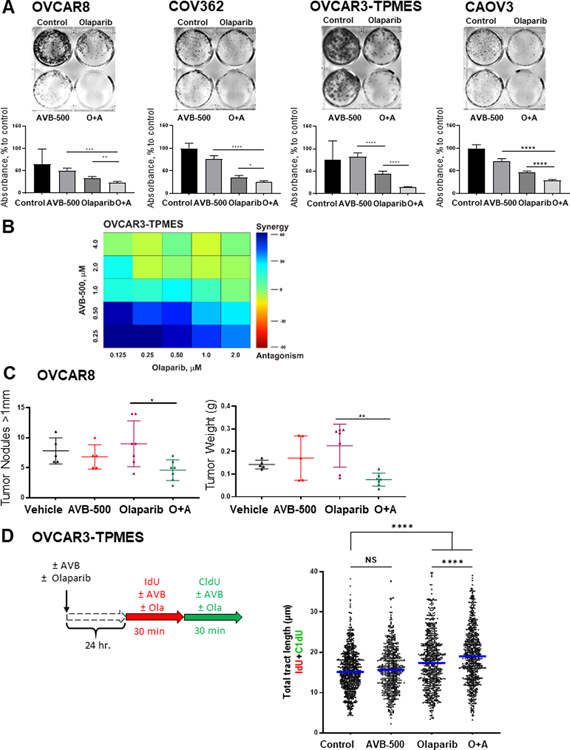
Figure 6. AXL inhibitor AVB-500 improves response to olaparib in vitro and in vivo.
A, Colony formation assay in cells treated with vehicle, 2μM Olaparib, 1μM AVB-500, or olaparib plus AVB-500 (O+A). Top, representative images; bottom, quantitation of percent to control absorbance at 590nm. Cells were treated for 72 hours and then incubated in media with 10% FBS until vehicle-treated cells formed colonies optimal for visualization. B, Loewe synergism analysis of OVCAR3-TPMES cells treated with varying doses of olaparib and AVB-500. C, Tumor burden of mice intraperitoneally injected with OVCAR8 as quantified by tumor nodule number and weight after treatment with vehicle, olaparib, AVB-500, or O+A. D, Schematic for DNA fiber assays. Total tract length of IdU+CldU in OVCAR3-TPMES cells treated with vehicle, 1 μM AVB-500, 10 μM olaparib, or O+A. Horizontal lines indicate medians. Cells were treated with 1 μM AVB-500 and 10 μM olaparib 24 hours before the start of the fiber assay. AVB-500 and olaparib were maintained in the cell media during the entire labeling period. **** P < 0.0001.

