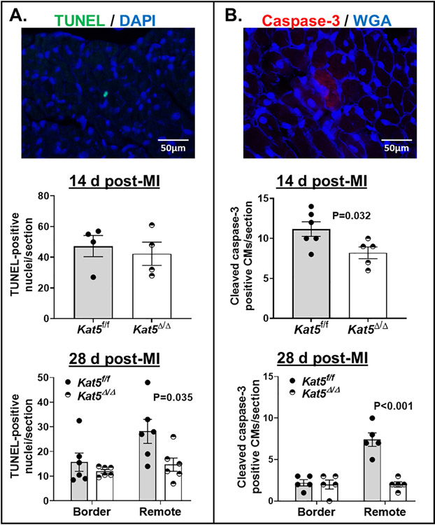
Figure 6. Reduced numbers of TUNEL- and caspase-3-positive cells in the infarcted Tip60-depleted hearts at days 14 and 28 post-MI.
Panel A. TUNEL/WGA-stained sections. Upper: representative 600x image. Lower: total number of TUNEL-positive nuclei per section; cell types were not identifiable. 14 d post-MI: N=4 mice per group; 28d post-MI: N=6 mice per group. Panel B. Caspase-3/WGA-stained sections. Upper: representative 600x image. Lower: total numbers of caspase-3-positive CMs per section; CM identity was inferred from cellular size based on WGA outline. N=5 mice for each time point per group. Data were reported as Mean ± SEM and compared by unpaired, two-tailed Student’s t tests with Welch’s correction.

