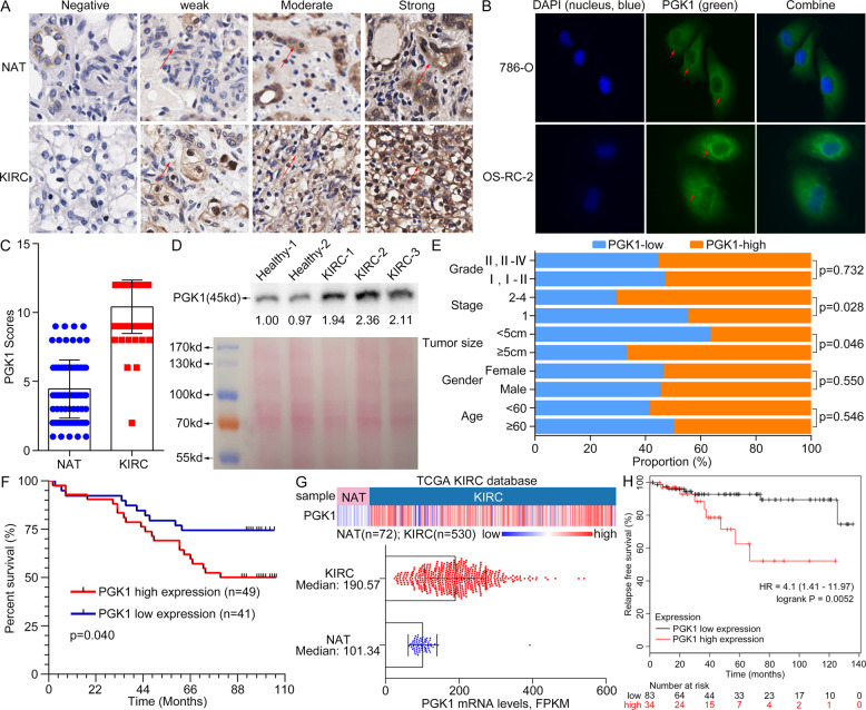
Fig. 4. PGK1 upregulation in tumor tissues and serum is indicative of poor prognosis for KIRC patients.
A Different staining level for PGK1 protein was shown in a paired noncancerous/tumor tissue from a commercial tissue microarray. Red arrows indicated positive staining for PGK1. B Cell endogenous PGK1 was mainly located in cytoplasm by cell fluorescence analysis. Red arrows meant positive fluorescent staining. C PGK1 level was increased in KIRC tissues by quantitative analysis of 90 cases of NATs and KIRC tissues. D The serum PGK1 was increased in KIRC patients compared to healthy persons by Western blot detection. Reversible Ponceau staining was used as a total loading control. E Associations of PGK1 levels and pathologic variables including grade, stage, tumor size, gender, and age obtained from tissue microarray. PGK1 protein level is well correlated with tumor size and tumor stage of KIRC (P < 0.05). F Kaplan–Meier overall survival curves for KIRC patients based on PGK1 expression from tissue microarray results. G Heatmap (top chart) and boxplot (bottom chart) for the transcriptional profiling of PGK1 in KIRC tissues (n = 530) and NATs (n = 72) respectively, derived from TCGA-KIRC species. H KIRC patients with high PGK1 mRNA level showed a shorter relapse-free survival (P < 0.05) from the KM-plotter database analysis.

