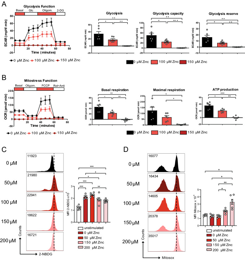
Figure 3.
Zinc aspartate reduces glycolysis and OXPHOS in CD4+ T cells. (A) Extracellular acidification rate (ECAR) of OT-II TCR tg CD4+ T cells in the absence and presence of 100 and 150 μM zinc aspartate after 24 h of stimulation and adding glucose, oligomycin, and 2-DG. Bar graphs show glycolysis, glycolysis capacity, and glycolysis reserve. (B) Oxygen consumption rate (OCR) of OT-II TCR tg CD4+ T cells in the absence and presence of 100 and 150 μM zinc aspartate after 24 h of stimulation and adding oligomycin, FCCP, and rotenone/antimycin. Bar graphs show basal respiration, maximal respiration and ATP production. (C) Glucose uptake determined using 2-NBDG and measured by FACS for the indicated groups. (D) Mitochondrial ROS production determined using Mitosox and measured by FACS for the indicated groups. Statistical analysis in (A–D) by one-way ANOVA followed by Tukey’s honestly significant difference (HSD) post hoc test for multiple comparisons. Data in (A–D) are from 3 experiments. Dots represent individual mice used to isolate OT-II TCR tg CD4+ T cells. *p < 0.05, **p < 0.01, ***p < 0.001.

