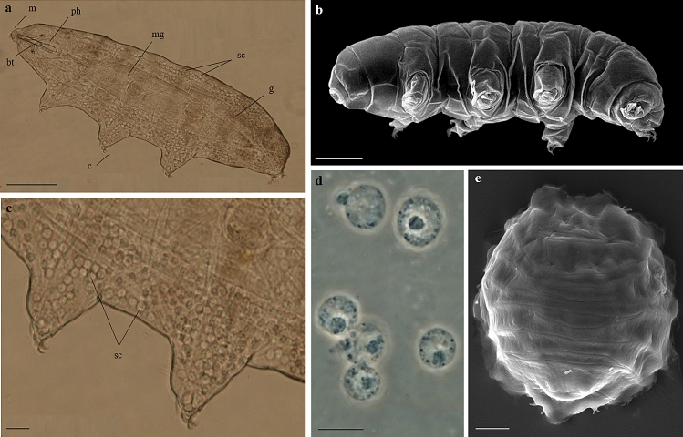
Figure 1.
Paramacrobiotus spatialis. (a) Specimen in toto and in vivo. (b) Specimen in toto. (c) Magnification of image (a) showing storage cells in the body cavity in correspondence to the second and third pair of legs. (d) Storage cells in vivo. (e) Desiccated specimen (tun). (a,c,d) LM (PhC), (b,e) SEM. Scale bars: a = 100 µm, b = 50 µm, c, e = 20 µm, d = 10 µm. bt = buccal tube; c = claws; m = mouth; mg = midgut; g = gonad; ph = pharynx; sc = storage cells.

