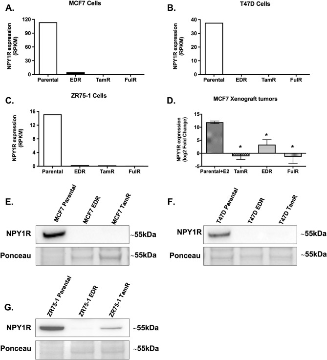
Figure 4.
NPY1R gene and protein expression is impaired in endocrine-resistant derivatives of ER+ BC cells in vitro and downregulated in tamoxifen-resistant xenograft tumors in vivo. RNA-Seq data of parental and their estrogen deprivation-resistant (EDR), tamoxifen-resistant (TamR), and fulvestrant-resistant (FulR) derivatives of (A) MCF7, (B) T47D, and (C) ZR75-1 cells were interrogated38. Data were plotted as Reads Per Kilobase of transcript per Million mapped reads (RPKM). (D) U133plus2 Affymetrix Chip array data from MCF-7 tumors derivatives of EDR, TamR, and FulR, alongside parental tumors in presence of continued estrogen supplementation (E2) was evaluated and plotted as RNA Express-calculated signal intensity (log2 transformed fold change)36,37. *indicates p < 0.05 by One-way ANOVA, Dunnett’s test (n = 3–4). The protein expression of NPY1R was compared in MCF7, T47D and ZR75-1 parental cells to their EDR and TamR derivatives. Representative immunoblotting images showing the protein expression of NPY1R in (E) MCF7, (F) T47D and (G) ZR75-1 parental cells and their EDR and TamR derivatives. Ponceau staining was used to assess equal protein loading for visual assessment. The densitometric quantitation of the relative intensity of NPY1R to parental cells (from three independent experiments) is shown in Supplementary Fig. 5. Full-length blots are presented in Supplementary Fig. 5.

