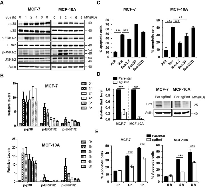
Fig. 1. p38 signaling and Bmf contribute to anoikis in mammary epithelial cells.
A MCF-7 or MCF-10A cells were trypsinized, resuspended and plated in poly-HEMA coated dishes and cultured for various time before lysed for western blot analysis. Attached cells were used as the “0 h” treatment groups. B Quantitation of the p-p38, p-ERK1/2 and p-JNK1/2 blots from (A), N = 3, error bars represent standard deviations. C MCF-7 or MCF-10A cells were replated in poly-HEMA coated dishes for 8 h in the presence or absence of indicated inhibitors (LY 2228820, 1 μM; AZD6244, 10 μM; SP 600125, 10 μM). Cells were then harvested for apoptosis analysis using annexin V-PI staining and flow cytometry. D qRT-PCR and western blot analysis of Bmf in MCF-7 and MCF-10A cells transduced with lentiviruses expressing Bmf sgRNA (sgBmf). E Parental or sgBmf lentivirus-transduced MCF-7/MCF-10A cells (polyclonal) were replated in poly-HEMA coated dish for 0, 4 and 8 h and assay for apoptosis using annexin V-PI staining and flow cytometry. All quantitative results were shown as mean ± STD from three independent experiments. Significance was determined by ANOVA one-way test, *p < 0.05; **p < 0.01, ***p < 0.001.

