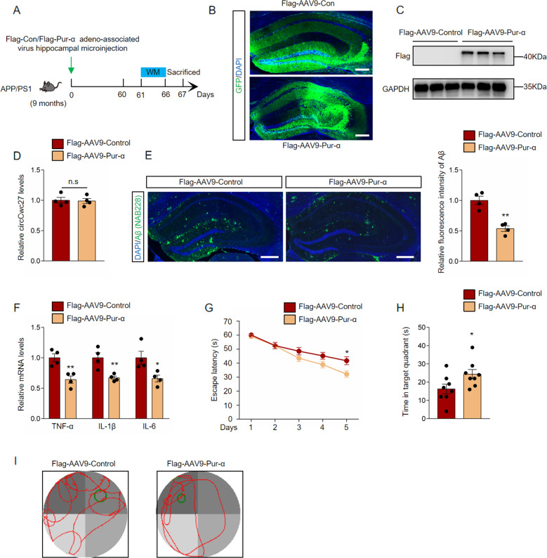
Fig. 5. Pur-α reduces Aβ deposition and prevents cognitive decline in AD.
A Timeline of experimental procedure in our research. B Confocal images of hippocampus were shown, and GFP (green) were used to visualize viral diffusion. Scale bar, 200 μm. C Flag levels were determined by immunoblotting in the hippocampus of APP/PS1 mice injected with Flag-AAV9-Control or Flag-AAV9-Pur-α. GAPDH was used as an inner control. n = 3. D qRT-PCR was used to detect the relative circCwc27 levels in the hippocampus of Flag-AAV9-Control or Flag-AAV9-Pur-α injected mice. n = 4. E Confocal images indicating DAPI (blue) for nuclei, NAB228 (green) for an amyloid plaque in the hippocampus from the APP/PS1 mice injected with Flag-AAV9-Control or Flag-AAV9-Pur-α. Scar bar: 200 μm. Amyloid plaque deposition was quantified and normalized to Flag-AAV9-Control group. n = 4. **P < 0.001 versus APP/PS1-Flag-AAV9-Control group using Student’s t-test. F qRT-PCR was used to detect cytokines levels in the hippocampus of APP/PS1-Flag-AAV9-Control or APP/PS1-Flag-AAV9-Pur-α injected mice. n = 4. *P < 0.05, **P < 0.01 versus APP/PS1-Flag-AAV9-Control group using Student’s t-test. G Latency to escape to a hidden platform in the MWM task during a 5-day training phase. Day 5: APP/PS1-Flag-AAV9-Pur-α (n = 8) versus APP/PS1-Flag-AAV9-control group (n = 8); *P < 0.05. Data are analyzed with Student’s t-test. H Probe trial was performed 24 h after the last training day and the time spent in the quadrant containing the hidden platform was recorded. n = 8. *P < 0.05 versus APP/PS1-Flag-AAV9-Control group using Student’s t-test. I Representative swimming trajectories from different groups of mice were shown. The green circle represented the hidden platform. All data in the figure are shown as mean ± SEM.

