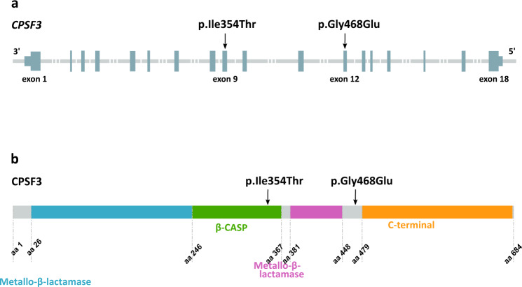
Fig. 1. Structure of CPSF3 (RefSeq transcript NM_016207.3) and location of the two missense variants found in homozygous state in patients with a severe intellectual disability syndrome.
a The p.Ile354Thr and p.Gly468Glu variants are located in exon 9 and 12 of CPSF3, respectively. Dark gray sections represent exons and untranslated regions, light gray lines represent introns. b The p.Ile354Thr variant is in the β-CASP domain of CPSF3 (amino acids 246–367, shown in green). The p.Gly468Glu variant is located within an uncharacterized linker domain (amino acids 448–479) between the critical second metallo-β-lactamase domain (shown in pink) and the highly conserved C-terminal domain (amino acids 479–682, shown in yellow) of CPSF3. The first metallo-β-lactamase domain is shown in blue, uncharacterized domains are shown in gray.

