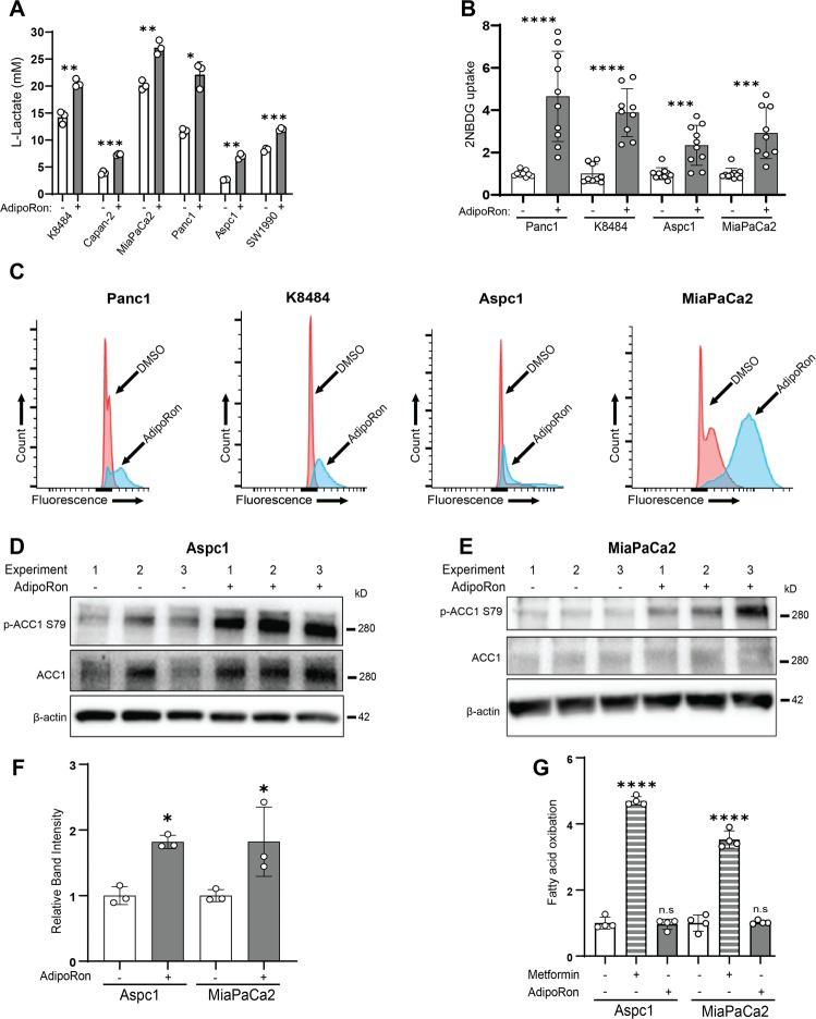
Fig. 4. AdipoRon increases glucose utilization.
A PDAC cells were cultured in reduced serum (2.5% fetal bovine serum) conditions for 24 h prior to treatment with 25 μM AdipoRon for 48 h and assessed for levels of secreted lactate (n = 3). B, C PDAC cells were treated with AdipoRon for 48 h, and then glucose- and serum-starved for 40 min prior to incubation with 2NBDG (200 µM) for 80 min. At the endpoints, the cells were washed and assessed for 2NBDG content either by plate reading (excitation 475 nm, emission 550 nm) or flow-cytometric analysis (B, n = 10; C, n = 4). D–F Levels of phospho-ACC1 were assessed in response to AdipoRon treatment (6 h), blots were cropped, and levels adjusted for clarity (n = 3). G Fatty acid oxidation was measured following AdipoRon treatment (6 h), n = 4. Statistics: t-test (F) or one-way ANOVA (G); error bars, SD; *P < 0.05.

