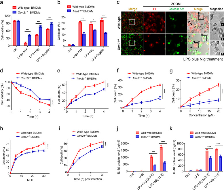
Fig. 4. Depletion of TRIM21 alleviates pyroptosis.
a, b Primary BMDM cells were isolated from WT and Trim21-/- C57BL/6 mice. Cells were primed with 1 μg/mL LPS for 2 h followed by 5 mM ATP or 20 μM Nig or 30 μM flagellin for 30 min. Cell viability (a) and cell death (b) rates were determined by the Cell Counting Kit-8 (Beyotime) and the LDH Cytotoxicity Assay Kit (Promega), respectively. c The alive WT and Trim21−/− iBMDM cells were stained by the green calcein, while dead cells were stained by the red PI. Representative data are shown from cells exposed to LPS plus Nig. Arrows indicate the bubbling of pyroptotic cells. Scale bar, 25 μm. d–f Time-course effects of TRIM21 on protecting iBMDM cells or primary BMDM cells against 1 μg/mL LPS plus 20 μM Nig-induced pyroptosis. g WT and Trim21−/− BMDM cells were primed with LPS (1 μg/mL) and treated with Nig at the indicated doses, followed by cell death analysis. h Effects of increased concentrations of S. Typhimurium (MOI = 0–30) on the cell death after 3 h exposure. i WT and Trim21−/− BMDM cells were infected by S. Typhimurium (MOI = 10) at different time points, followed by cell death analysis. j, k WT and Trim21−/− BMDM cells were primed with LPS (1 μg/mL) and treated with 20 μM Nig at the indicated times, followed by cytokine secretion measurement. The data are the means ± SD of triplicate samples from a representative experiment (*P < 0.05; **P < 0.01; ***P < 0.001; ****P < 0.0001). All data are representative of three independent experiments.

