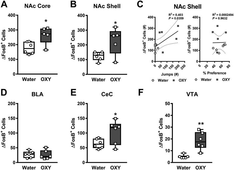
Fig. 7. Naloxone challenge following oral oxycodone consumption leads to greater numbers of ΔFosB expressing cells in the NAc core, shell, CeC, and VTA.

Following naloxone challenge, oxycodone consuming mice showed higher numbers of ΔFosB expressing cells in the A) NAc core, B) NAc shell, E) CeC, and F) VTA but not the D) BLA when compared to water consuming mice. C) The number of naloxone-precipitated withdrawal jumps was positively correlated with the number of ΔFosB-expressing cells in the NAc shell (left panel). In contrast, no significant correlations was observed between the % preference and the number of ΔFosB-expressing cells in the NAc shell (right panel). (Nucleus accumbens – NAc, Basolateral amygdalar nucleus - BLA, Central amygdalar nucleus capsular part – CeC, Ventral tegmental area - VTA) Data are expressed as mean ± S.E.M. (n = 5-6 per group). *p < 0.05 vs. water consuming mice.
