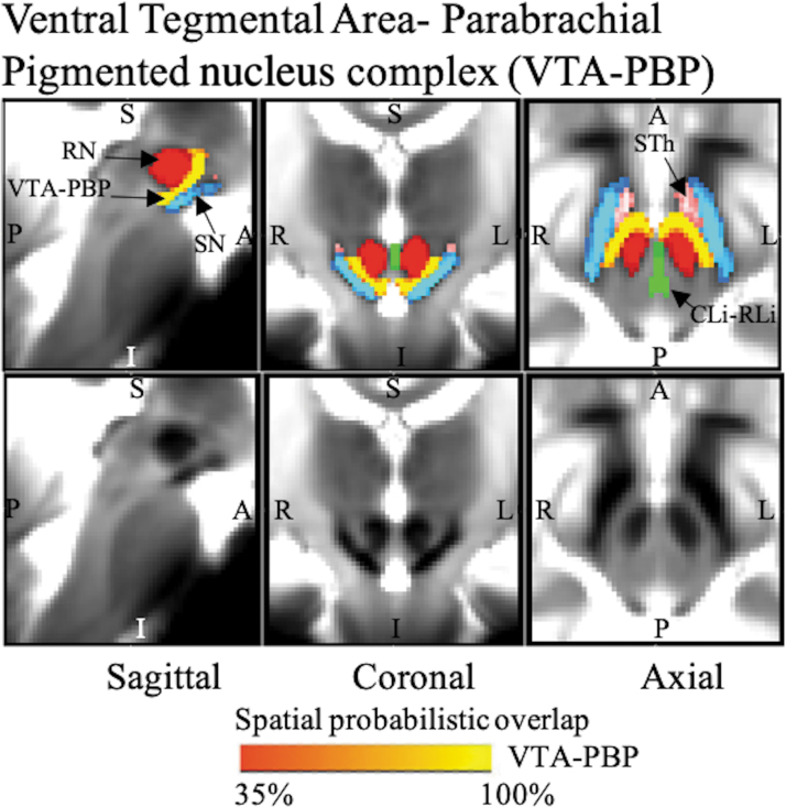
FIG. 4.
The probabilistic (n = 12) atlas label in the MNI space of VTA-PBP is shown (red-to-yellow) with neighboring nuclei (black arrows), such as STh (pink), SN (blue-light blue), RN (red), and CLi-RLi (green). As a background image, we used the T2w map in the MNI space. Very good (i.e., up to 100%) spatial agreement of labels across subjects was observed, indicating the feasibility of delineating the probabilistic label of this nucleus complex. Of note, we found good contrast for these nuclei, sandwiched between RN and SN, showing a horseshoe-shaped structure in the coronal and axial sections, which matched the description of these nuclei from literature (Olszewski and Baxter, 1954; Paxinos et al., 2012). CLi-RLi, caudal–rostral linear nucleus of the raphe; RN, red nucleus; SN, substantia nigra; STh, subthalamic nucleus; VTA-PBP, ventral tegmental area–parabrachial pigmented nucleus complex.

