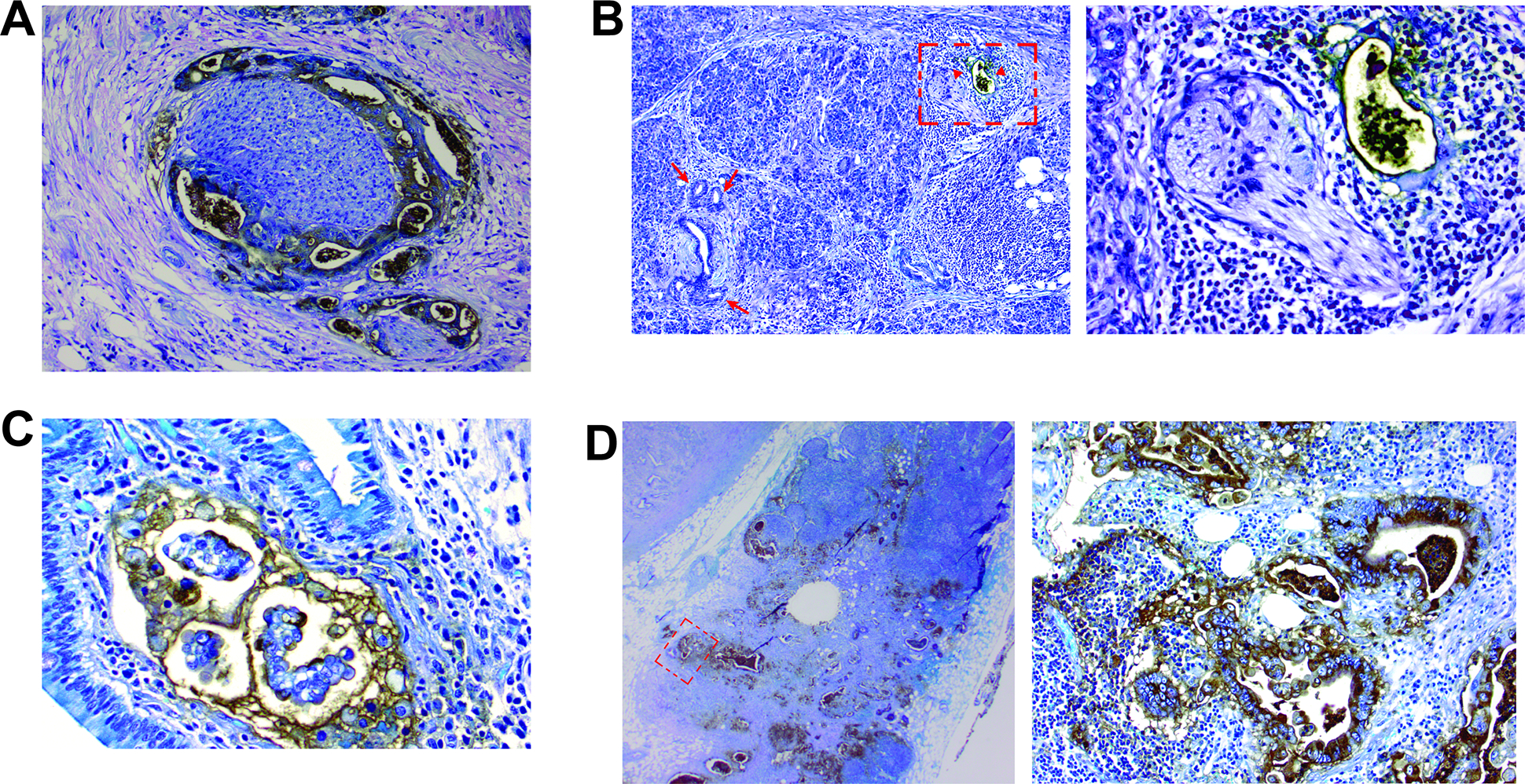
Figure 4:

A) Das-1 staining highlighting perineural invasion. B) A malignant duct in the vicinity of neural tissue is positive for Das-1 (arrow heads; an area marked by a rectangle is magnified in the right-side image), while normal interlobular duct and low-grade PanIN lesions are non-reactive to Das-1 (arrows). C) Lymphatic invasion with Das-1 expression. D) An example of lymph node metastasis with grade 3 Das-1 expression (an area marked by a rectangle is magnified in the right-side image).
