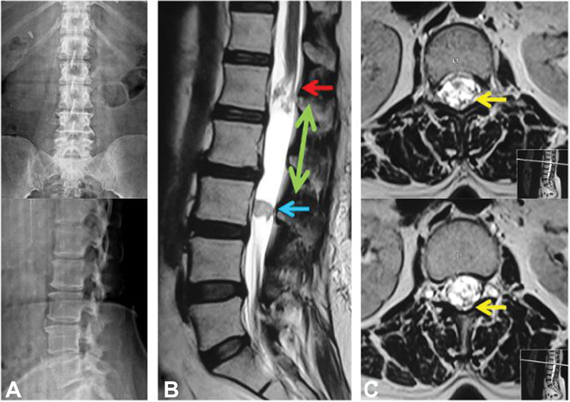
Fig. 1.

( A ) Plain radiography revealed lumbar spondylotic changes in the form of anterior disc osteophytes at L3–L4 and L4–L5 level with no evidence of instability. ( B ) Sagittal T2-weighted image shows an elongated predominantly cystic (green arrow) intradural-extramedullary lesion extending from the lower endplate of L1 to L3 vertebra. There is a solid intramural homogeneous nodule measuring 1.6 × 1.6 cm (blue arrow) at the level of the mid-L3 vertebral body. Conus is distorted with splaying and clumping of the nerve roots (red arrow) ( C ) Axial T2-weighted images showing a few prominent flow voids at the level of conus (yellow arrows).
