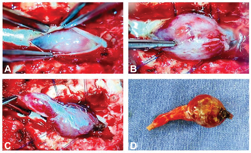
Fig. 4.

( A ) Postdurotomy surgical microscope picture before delineating the tumor. ( B ) Large tumor bulging out from the canal. ( C ) Both solid and cystic components of the tumor are very well-appreciable with a leash of vessels on the surface as well as at the poles of the tumor after delineating the tumor margins and ( D ) postexcision lesion appears shrunken in size due to decongestion.
