Figure 1.

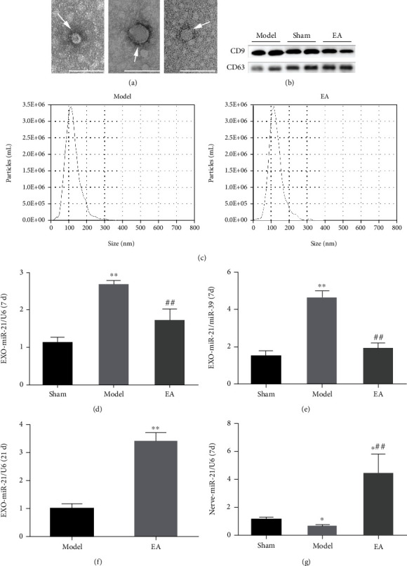
EA regulated the release of serum exosomal miR-21. (a) Exosomes (arrows) were observed under a transmission electron microscope, bar = 100 nm. (b) The surface markers of the exosomes, including CD9 and CD63, were detected using WB. (c) The particle size was detected using NTA. RT-qPCR was conducted to detect the relative expression level of miR-21 in the serum exosomes of rats in each group at 7 d with U6 used as the internal reference (d) or miR-39 used as an external reference (e). ∗∗P < 0.01 compared with the sham group; ##P < 0.01 compared with the model group. (f) RT-qPCR detection of miR-21 expression in serum exosomes at 21 d. ∗∗P < 0.01 compared with the model group. (g) RT-qPCR detection of the relative expression of miR-21 in locally injured nerves of rats at 7 d. ∗P < 0.05 compared with the sham group; ##P < 0.01 compared with the model group.
