Figure 10.

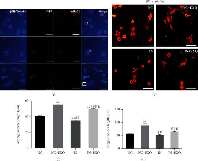
SC-derived exosomal miR-21 promotes neurite outgrowth in vitro. (a) miR-21 in situ hybridizations (red) and immunofluorescence staining of βIII-tubulin (blue) showed that exosomes secreted by SC could be taken up by neurons. miR-21 was found in the neuron cell body (indicated by arrows) and neuronal processes (shown in the square). Bar = 25 μm. (b) Immunofluorescence staining of βIII-tubulin (red) to detect NG108-15 protrusion growth. Bar = 50 μm. (c) The average protrusion length of the neurons were calculated. ∗∗P < 0.01 compared with the NC group; ##P < 0.01 compared with the NC + EXO group; &&P < 0.01 compared with the IN group. (d) The length of the longest protrusion was calculated. ∗∗P < 0.01 compared with the NC group; ##P < 0.01 compared with the NC + EXO group; &P < 0.05, compared with the IN group.
