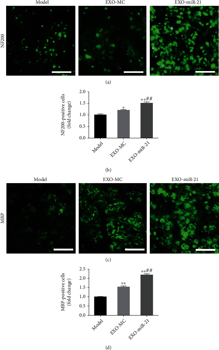
Figure 8.

Exosomal miR-21 participates in the regeneration of nerve fibers. (a) Immunofluorescence staining of NF200 (green). Bar = 10 μm. (b) The number of NF200 positive cells, which was used to indicate the number of regenerated axons, was counted. ∗P < 0.05; ∗∗P < 0.01 compared with the model group; ##P < 0.01 compared with the EXO-MC group. (c) Immunofluorescence staining of MBP (green). Bar = 10 μm. (d) Statistics of the number of MBP positive cells, which was used to indicate the number of myelin sheaths. ∗∗P < 0.01 compared with the model group; ##P < 0.01 compared with the EXO-MC group.
