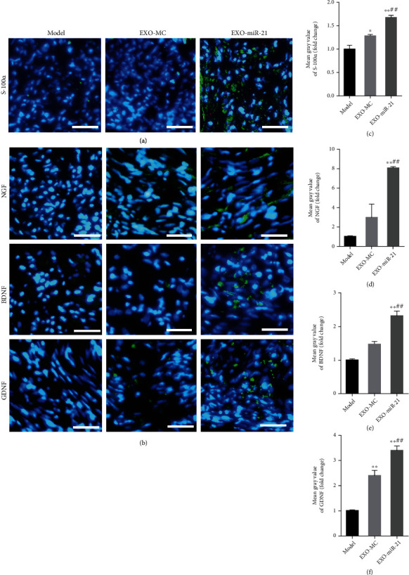
Figure 9.

Exosomal miR-21 participates in the proliferation of SC and the expression of NTFs. (a) Immunofluorescence staining of S-100α (green). The nuclei were visualized using DAPI (blue) staining. Bar = 10 μm. (b) Immunofluorescence staining of NGF, BDNF, and GDNF (green). Bar = 10 μm. (c) Mean fluorescence intensity of S-100α. ∗P < 0.05; ∗∗P < 0.01 compared with the model group; ##P < 0.01 compared with the EXO-MC group. Mean fluorescence intensity of NGF, BDNF, and GDNF (d–f). ∗∗P < 0.01 compared with the model group; ##P < 0.01 compared with the EXO-MC group.
