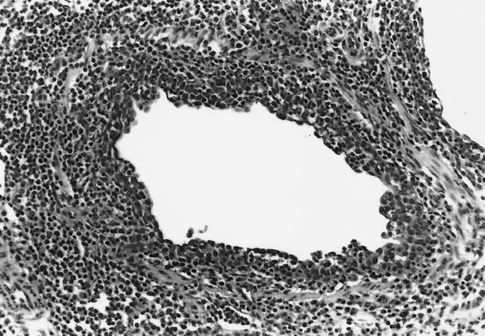
FIG. 3.
Bronchiole in the lung of a pig inoculated with both M. hyopneumoniae and SIV. The pig was euthanatized 3 DPI with SIV and 24 DPI with M. hyopneumoniae. The bronchiole exhibits the characteristic disruption and attenuation of the epithelial layer and early irregular reactive proliferation subsequent to SIV infection and intense peribronchiolar lymphocyte infiltration induced by both virus and mycoplasma.

