
Figure 3. Sequence features of effector domains.
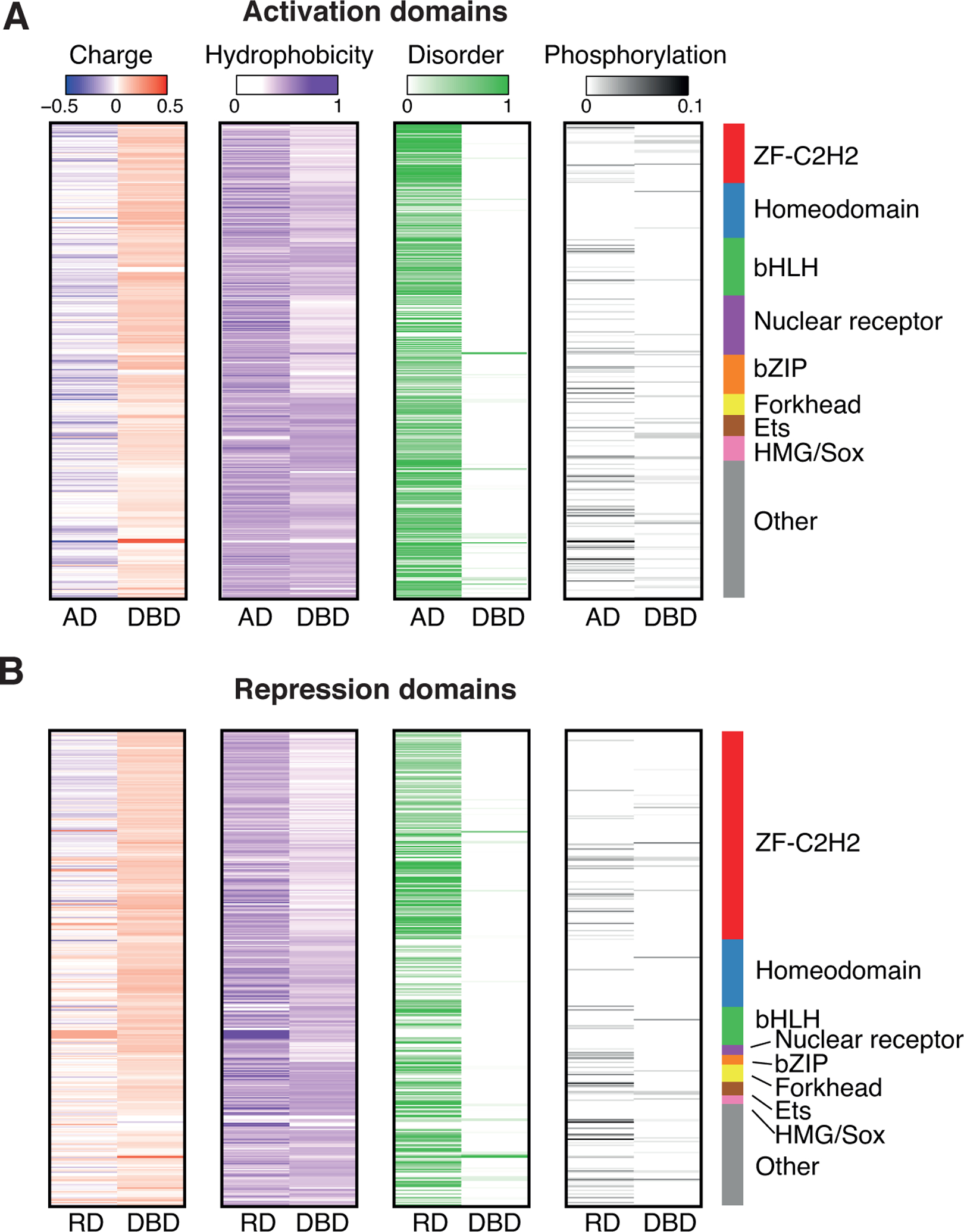
(A-B) For each activation domain (A) and repression domain (B) the charge density (charge / amino acid length), hydrophobicity, disorder (determined using AlphaFold), and phosphorylation density (number of phosphorylation events / amino acid length) are indicated.
See also Figures S2 and S3.