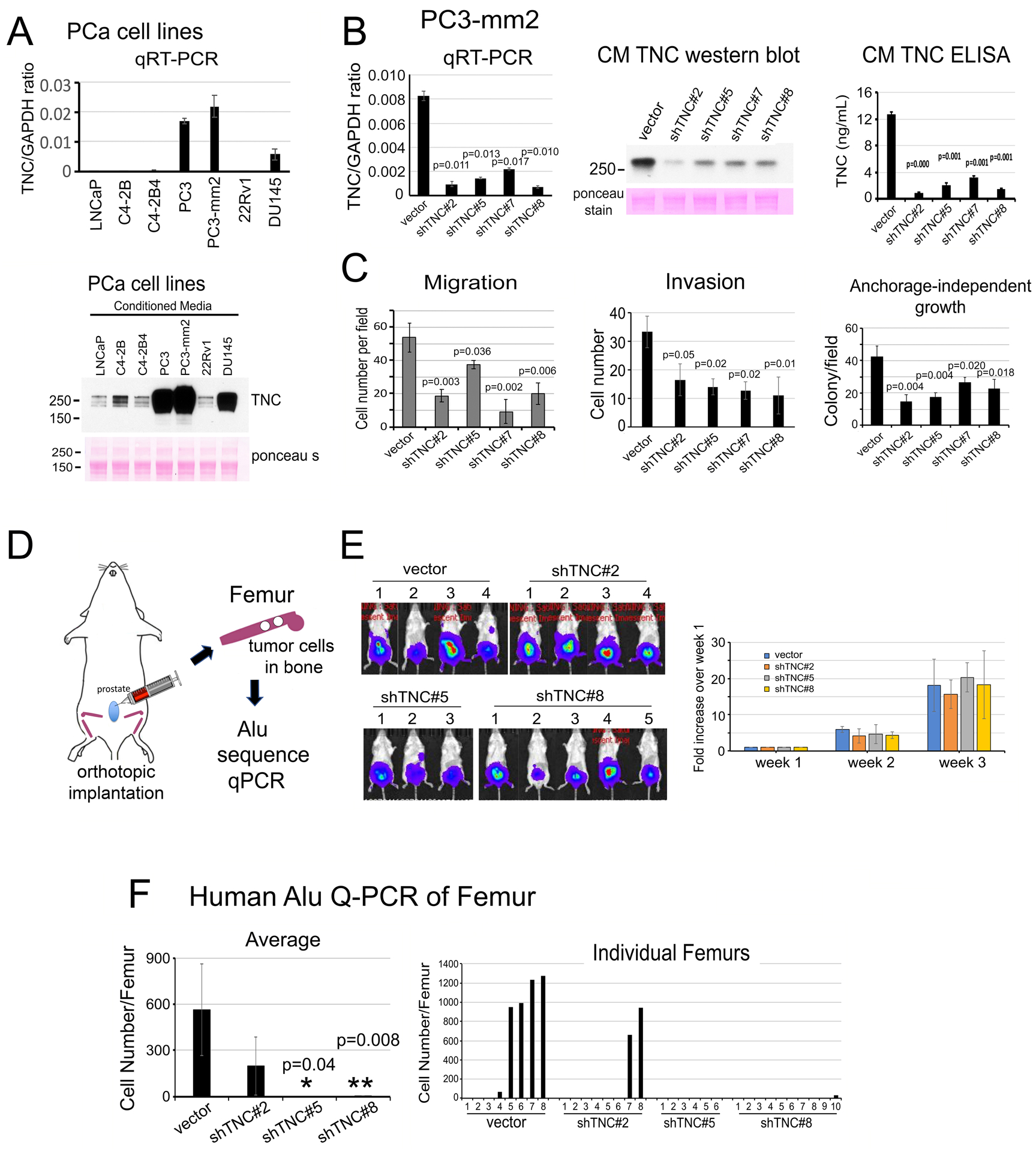
Figure 5. Knockdown of Tenascin C decreases the migration, invasion, anchorage-independent growth of PC3-mm2 cells in vitro and the metastasis of PC3-mm2 cells to bone in vivo.

(A) qRT-PCR for TNC mRNA in PCa cell lines (upper). Western blot of TNC protein in CM from PCa cell lines (lower). (B) qRT-PCR (left) for TNC mRNA in PC3-mm2-shTNC#2, #5, #7 and #8 clones. CMs from PC3-mm2-shTNC clones were analyzed by Western blot (middle) and ELISA (right). (C) Migration, invasion, and anchorage-independent growth of PC3-mm2-shTNC clones in (B). (D) Experimental scheme. PC3-vector, shTNC#2, #5, or #8 cells were injected orthotopically into mouse prostate. After 3 weeks, femurs were dissected and total DNA prepared. Tumor cells that metastasized to bone were determined by human Alu sequence qPCR. (E) Bioluminescence of tumors in the various mouse groups at 3 weeks post-injection. Time course of tumor growth based on bioluminescence is shown. (F) Quantification of tumor cells that have metastasized to bone. Left, average number of tumor cells metastasized from prostate to bone. Right, number of tumor cells detected in individual legs in control mice and PC3-mm2-shTNC#2, shTNC#5, or shTNC#8 injected mice. P values were by Student’s t-test.
