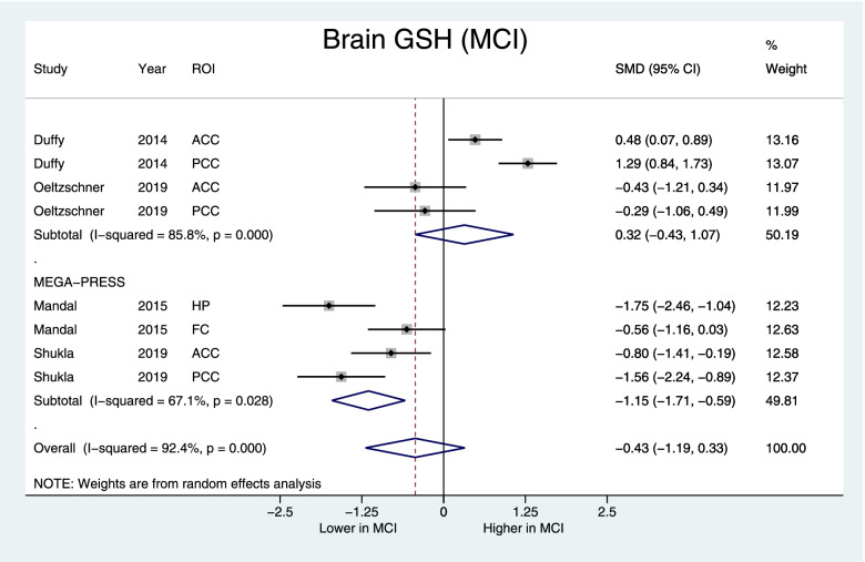
Fig. 4.
Forest plot displaying brain GSH concentrations in MCI and control subjects, with the subgroup of studies using the MEGA-PRESS protocol at the bottom. Shown are the standardized mean differences (SMD) and 95% confidence intervals (95% CI). Negative values denote lower GSH in MCI subjects while positive values denote higher in GSH in MCI compared to controls. Pooled SMD [95% CI] = −0.43 [−1.19, 0.33], z=1.12, p=0.26, MEGA-PRESS subgroup: SMD [95% CI] = −1.15 [−1.71, −0.59], z=4.0, p<0.001. ROI indicates the region of interest: ACC anterior cingulate cortex, PCC posterior cingulate cortex, FC frontal cortex, HP hippocampus

