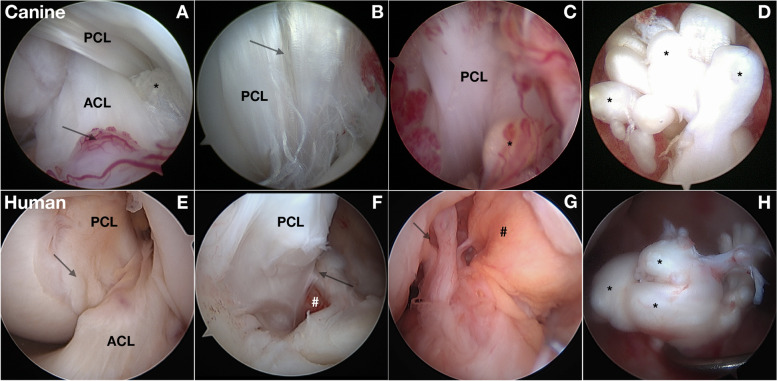
Fig. 4.
Intraarticular findings associated with anterior cruciate ligament (ACL) rupture in the dog and human. A-D Arthroscopic views of the intercondylar notch (ICN) in dogs with anterior cruciate ligament rupture (ACL) (*). A Fiber rupture often involves specific fiber bundles in the anteromedial bundle of the ACL (*). Associated synovitis is present (arrow). B Fiber rupture and splitting (arrow) of the posterior cruciate ligament (PCL) is also common. C With progressive fiber rupture, associated synovitis reflects hypertrophy, vascularity and inflammatory changes. The healing response in fiber bundles (*) is not successful. D View of the tibial attachment of a complete ACL rupture. A marked healing response in ruptured fiber bundles (*) leads to enlargement of ruptured fiber bundles. E-H Arthroscopic views of a human knee with ACL rupture. E The femoral ICN containing both ACL and PCL as they twist around each other with overlying synovium (arrow) is similar to dogs. Both species develop an associated synovial inflammatory response. F PCL fiber rupture (arrow) with adjacent synovitis and hemorrhage (#). G The ACL rupture can be seen with few fibers remaining attached to the femur (arrow) with synovitis (#) overlying the PCL. H The blunted end of ruptured ACL fibers at the tibial attachment, similar to the dog

