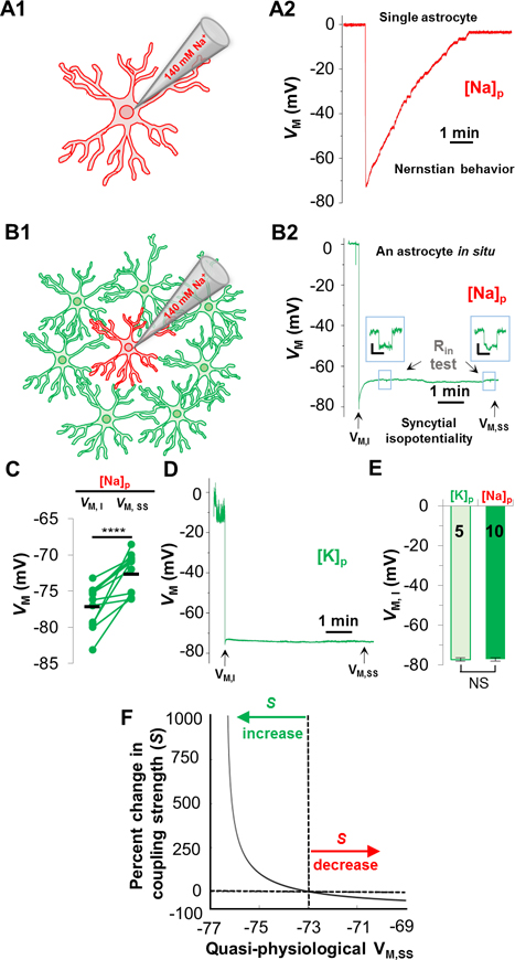
Figure 1. Astrocyte syncytial isopotentiality.
(A) In a single freshly disassociated astrocyte, K+-free/Na+ pipette solution [Na+]P progressively substituted the endogenous K+ content in whole-cell recording, leading to a VM depolarization towards 0 mV as predicted by Nernstian equation. (B) The VM recorded from an astrocyte in situ with [Na+]P disobeys the Nernstian prediction. Instead, the VM maintains at a quasi-physiological level. (B2) In [Na+]P recording, the initial VM (VM,I), and the quasi-physiological VM at the steady-state (VM,SS). Input resistance (RIn) was periodically monitored during recording. (C) VM,SS was significantly more depolarized than VM,I (p < 0.001, pairwise t-test). (D) VM,I and VM,SS was measured in whole-cell recording using 140 mM KCl recording pipette ([K+]P). (E) VM,I in [K+]P and [Na+]P recordings was not significantly different (p > 0.05, unpaired t-test). (F) The relationship between VM,SS and coupling strength (S) predicted by computational modeling. The VM,SS from hippocampal CA1 syncytium, −73 mV, is used as the baseline in the model to predict the relative difference in coupling strength (S%) between CA1 syncytium and a syncytium of another brain region.

