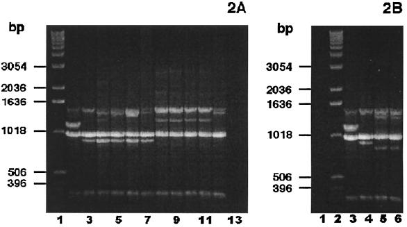
FIG. 2.
P. multocida subsp. multocida PCR fingerprint analyses. (A) Nine sorbitol-positive, α-Glu-negative P. multocida subsp. multocida bite wound isolates, examined by PCR fingerprint analyses using the single primer, M13 core, displayed either the group II or group III PCR fingerprint profile. (Sorbitol fermentation was based on a consensus of API-20E, PRAS, and Andrades sorbitol fermentation reactions.) Lane 1, DNA ladder; lane 2, P. multocida subsp. septica ATCC 51688; lane 3, P. multocida subsp. multocida ATCC 12947; lanes 4 to 7, bite wound isolates demonstrating the group II PCR fingerprint pattern; lanes 8 to 12, bite wound isolates showing the group III PCR fingerprint profile; lane 13, negative control (no DNA template). (B) Two sorbitol-positive, α-Glu-positive P. multocida bite wound isolates, examined by PCR fingerprinting analyses using the single primer, M13 core, exhibited the group III PCR fingerprint profile characteristic of other P. multocida subsp. multocida clinical isolates. Lane 1, negative control (no DNA template); lane 2, DNA ladder; lane 3, P. multocida subsp. septica ATCC 51688; lane 4, P. multocida subsp. multocida ATCC 12947; lanes 5 and 6, bite wound isolates.

