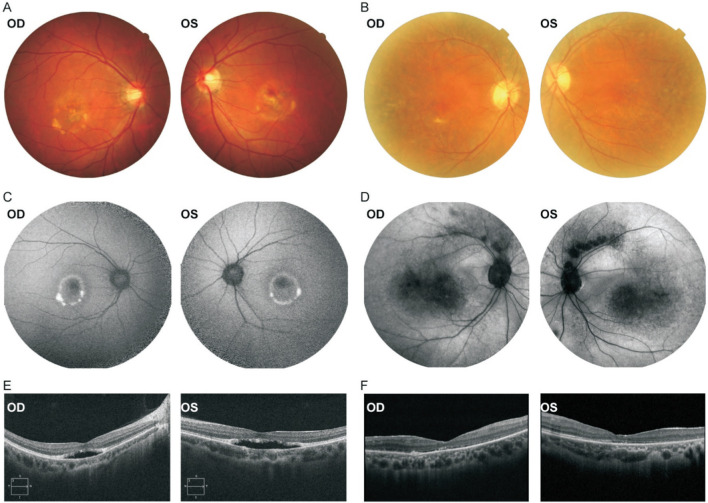
Figure 1. Retinal images and SD-OCT images of Family 1.
The color fundus photographs reveal bilateral macular yellow vitelliform lesions in the proband (A) and bilateral peripheral retinal mottled pigmentary changes in his mother (B). The FAF images reveal the central hypoautofluorescence surrounded by an annulus of inhomogeneous hyperautofluorescence in each eye in the proband (C) and reveal hypoautofluorescent blocks within the macular area and along the supratemporal vascular arcade in his mother (D). The SD-OCT scans show marked serous macular detachment in the central macula in each eye in the proband (E) and slight serous macular detachment in his mother (F). OD: Right eye; OS: Left eye.

